Robert M. Bernstein, MD,* and William R. Rassman, MD
Dermatologic Clinics 1999; 17 (2): 277-95.
*Assistant Clinical Professor of Dermatology, College of Physicians and Surgeons,
Columbia University, New York, NY
From the College of Physicians and Surgeons, Columbia University, New York, New York (RMB) and the New Hair Institute, New York, New York (RMB and WRR)
Follicular Unit Transplantation is a method of hair restoration surgery where hair is transplanted exclusively in its naturally occurring, individual follicular units. The evolution and rational for follicular unit transplantation will be discussed, as well as the logic for the various techniques used in its implementation. Specifically, the logic for single strip harvesting, stereo-microscopic dissection, automated graft insertion, and large transplant sessions will be reviewed. The central role of the follicular unit constant in the surgical planning will also be discussed.
Progress in modern medicine is often the result of sophisticated technology that allows us to quietly probe into the deepest regions of the human body or to analyze its actions at the molecular level. Rarely, is a field of medicine dramatically changed by simple observation. After more than three decades of relative inertia, surgical hair restoration is undergoing such a change. This writing discusses the logical applications of these observations to clinical practice, a logic that has literally revolutionized hair transplantation in just a few short years. It will also touch upon some of the illogical judgments that contributed to its delay.
HISTORICAL ASPECTS
“A donor (graft) is better if it is as small as possible. The reason is that if a donor is big, hairs grow in … a very unnatural appearance.” ~ Hajime Tamura – 1943
If we had only heeded the advice of the pioneering Japanese hair transplant surgeons in the first half of this century, we could have avoided years of unsightly surgical results that caused dismay to thousands of unwary patients, and literally tarnished an entire field of medicine. Unfortunately, the “Japanese insight” was lost to us during World War II and when we tried to “re-invent the wheel,” we did it wrong.
The Punch Graft Technique
After the “rediscovery” of hair transplantation by Dr. Norman Orentreich in 1952, the excitement that hair actually grew, and continued to grow after it was transplanted, clouded the very essence of hair restoration surgery i.e., that it was a cosmetic procedure whose sole purpose was to improve the appearance of the balding patient. The 4-mm plug that had been ordained as the optimal vehicle for moving hair was actually of a size that had no counterpart in nature.
The initial problem was that the decision to use 4-mm plugs was based mainly upon technical rather than aesthetic considerations. In the original, ingenious experiments performed by Dr. Orentreich, published in the Annals of the New York Academy of Science in 1959, which established the concept of “Donor Dominance,” 6 to 12-mm punches (trochars) were used to create the grafts.18 At these sizes, there was an unacceptably high rate of hair loss in the center of the grafts due to the difficulty of oxygen to diffuse over such large distances. The initial effort to decrease graft size was thwarted by the concern that much smaller grafts would not move enough hair to make the procedure worthwhile. Eventually a compromise was reached, and the 4mm graft was born.
In addition, a logic developed which postulated that by replacing bald skin with hair bearing skin, most of the balding area could eventually be replaced with hair. No adjustments for scar contraction were accounted for, and no changes in the size of the newly transplanted grafts were expected, despite observations to the contrary. More importantly, these assumptions were based upon the mathematically impossible feat of covering a large area of balding with a much smaller donor supply, while maintaining the same density.
The punch-graft, open-donor technique was developed with tools in routine use by dermatologists of the time. In the “open-donor method” devised by Dr. Orentreich, the same trochar that was used to make the recipient sites was also used to harvest the hair. Since hair in the donor area emerges from the scalp at rather acute angles, that varies in different regions, the physician was required to have the angle of the trochar exactly parallel to the angle of the hair. If there was even the slightest deviation from a perfectly parallel orientation, significant wastage of hair would occur from follicular transection. In fact, in many patients, so much transection would occur that the potentially “pluggy” appearance was reduced to a thinner look by the inadvertent reduction in the number of hairs per graft.
The hidden problem, of course, was that this harvesting technique reflected a grossly inefficient use of the donor supply, and patients often became depleted of donor hair long before the transplant process was completed. These problems were compounded by the fact that in the “open donor method” the wounds were left to heal by secondary intention and the resulting fibrosis further altered the direction of the remaining donor hair, making subsequent harvesting even more difficult.
The large donor and recipient wounds created by these punches necessitated that the procedure be performed in small sessions, usually 20 to 50 grafts at a sitting, with the sessions spaced apart in time due to the prolonged healing. As a result, one of the truly unfortunate problems intrinsic to the early techniques was that neither the long-term cosmetic issues, nor the ultimate depletion of the patient’s donor supply, could be appreciated for many years. Possibly because of Dr. Orentreich’s deservedly high esteem in the medical community (he also did pioneering work in dermabrasion, intra-lesional corticosteroids, injectable silicon, and the hormonal treatment of hair loss (to name just a few), the 4mm size went unchanged for years.
Early attempts at reducing the size of the grafts were largely unsuccessful. A reasonable approach to making these large plugs cosmetically more acceptable was to divide them into smaller grafts i.e. to produce split grafts or quarter grafts from the larger plugs.10 Unfortunately, these only resulted in further manipulation and damage to grafts that already contained populations of transected follicles. Simply reducing the size of the punches20 was also problematic since a decreased radius greatly exaggerated the damage caused by even the slightest deviation in the harvesting angle. It seemed that a relative impasse had been reached in trying to create a smaller size graft that would be cosmetically acceptable, contain enough hair to make the procedure worthwhile, and not be too wasteful of the donor supply. Fortunately, new techniques in harvesting the donor tissue provided solutions to these problems.
The Donor Supply
Some hair transplant surgeons, conscious of the finite donor supply, noticed that there were islands of hair bearing skin left behind after the initial rows of plugs were harvested. However, subsequent attempts to harvest the intervening tissue, and leaving the wounds open resulted in confluent areas of scarred scalp devoid of hair, and lacking adequate camouflage. Suturing the open donor wounds seemed to be a logical solution for decreasing the scarring, but this further altered the hair direction and made the remaining scalp less amenable to successful punch harvesting.
A more creative attempt to deal with this problem was to totally excise the tissue between the rows of punches and then suture the “serrated” upper edge to the serrated lower edge. The wound edges would then neatly come together if the punches were aligned properly.14,19 There were two important consequences of this procedure. The first was that it produced a piece of “free standing” donor tissue that could be cut into smaller pieces under direct visualization prior to transplantation. The second was that the sutured incision left a single line in the donor area (although somewhat squiggly). After looking at the result of this procedure even the casual observer would have to question the necessity of the punch graft aspect of the process. After all, why not just remove an intact strip of tissue and suture the wound edges closed, obviating the problems of the punches, i.e. the open donor wounds and the punch transection of hair follicles. After a number of years, this is the procedure that was eventually adopted.
The double- and then multi-bladed knifes11,24,28 were born out of an attempt to avoid the open donor scars produced by the punch-graft method, and to decrease the transection produced by this “blind” harvesting technique. Unfortunately, it solved only the first of these two problems. As with the punches, the multi-bladed knife was also a form of “blind-harvesting” since the surgeon would still have to match the angle of hair in order to avoid follicular transection, and the visual cues needed to perform this accurately were either hidden below the surface of the skin or covered with blood. As a result, the necessary fine-tuning of the blade angle, while making the incision, always came too late, i.e. after the transection of hair follicles had occurred.
In addition to the difficulty in following the curve of the skull, as one moved across the donor area horizontally, the fixed relationship between the multiple blades did not allow for any adjustments in the vertical plane. To compound the problem, pressure from one blade would distort the direction of hair near the others. The surgeon could adjust one blade (usually the upper) to follow the changing hair direction as he moved around the scalp, but since the angle of hair changed in the vertical dimension as well, transection caused by the other blades would be unavoidable. In addition, as one tried to angle the knife, in order to follow the vertical curve of the scalp, some blades might be too superficial, while others too deep. The superficial wound required a second incision which ran the risk of further transection. The deeper wound risked cutting fascia, or larger nerves and blood vessels, increasing the morbidity of the procedure.
An obvious solution would be to take a single strip of tissue from the donor area. The problem with that was that, in all but the smallest procedures, one was left with a large, three dimensional piece of tissue that defied further sectioning. In addition, with the trend to perform larger sessions, the fine slivers produced by the multi-blade knife grew more appealing, and the cumbersome nature of the single strip proved a hindrance to completing the hair transplant surgery in a reasonable period of time.
There was still one last important issue with the multi-bladed knife, i.e. that the blades moved in random planes though the donor tissue. As we will soon discuss, hair doesn’t grow randomly in the donor area, nor in the rest of the scalp for that matter, but in tightly organized bundles called follicular units. In effect then, the multi-bladed knife, even if it passed though the scalp perfectly aligned with, and parallel to, the growing hair, would still break up the integrity of these naturally occurring structures and reduce hair yield.
The significance of this last problem was not initially known, but it became apparent that transplanting very small grafts in large quantities, produced a thin look, and that this look was thinner than one would have anticipated based solely upon the amount of hair transplanted. The role of the multi-bladed knife in contributing to this problem is still in dispute, but it is felt by these authors, as well as other, to be very substantial. Other factors will be covered in later sections.
The recognition that square inch for square inch replacement of bald scalp with hair would not be possible with transplantation, posed a frustrating dilemma to the surgeon, and allowed a number of other procedures to proliferate, namely scalp reductions, lifts, and flaps. However, in the illusive goal of restoring original density, the mathematics of these procedures made no more sense than the plugs they were supposed to complement or replace. Regardless of the choice, the precious donor supply was still being consumed, and the patient’s long-term results compromised. In retrospect, it seems that the popularity of these procedures was not based upon their intrinsic value, but upon the fact that the alternatives were so poor. When the quality of the transplantation procedures finally improved, the frequency of these surgeries declined.
This is the position that the field of hair transplantation found itself in as it entered the 1990’s, and for the most part stayed in until 1996, when follicular unit transplantation caught on, and everything changed. As we will discuss, the logic of using follicular units is quite inseparable from the technique itself, and many of these techniques had their foundation as far back as 1982 in the “Punctiform” procedure of Carlos Uebel26 that later evolved into the “Megasession” and in the work of Dr. Bobby Limmer who began using microscopic dissection and single strip harvesting as far back as 1988.17 Surprisingly, the fact that hair grows in discrete bundles was largely unknown to most hair transplant surgeons for almost forty years, and it was the general revelation that these naturally occurring groups could be used to the patient’s advantage that changed the hair transplant procedure forever.
THE LOGIC OF PRESERVING THE FOLLICULAR UNIT
The underlying premise of follicular unit transplantation is that the intact, individual follicular unit is sacred. It should neither be broken up into smaller units, nor combined into larger ones.5,7,9 This simple idea may not seem like a radical approach to hair transplantation, but when viewed in the context of how the surgery has been performed over the past forty years (when the very existence of the follicular unit went generally unrecognized), it is radical indeed. Even now when its existence is widely known, there is a trend in hair transplantation to not only ignore the importance of the follicular unit, but to ignore the integrity of the follicle itself.
The follicular unit (see figure 1) was first defined by Headington in his landmark 1984 paper “Transverse Microscopic Anatomy of the Human Scalp.13 The follicular unit includes:
• 1 to 4 terminal follicles
• 1, or rarely 2, vellus follicles
• associated sebaceous lobules
• insertions of the arrector pili muscles
• perifollicular vascular plexus
• perifollicular neural net
• perifolliculum – cirumferential band of fine adventitial collagen that defines the unit
This rather dry definition belies the fact that the follicular unit is a physiologic entity rather than just an anatomic one. As we will see, the obvious reason to preserve the integrity of the follicular unit is economy of size, i.e. it is a way to get the most hair into the smallest possible site, and create the smallest wound. The ingenious hair transplant surgeon, Dr. David Seager, gave us another.23 In a bilateral controlled study, matched for the number of hairs, he showed that when single-hair micrografts were generated from breaking up larger follicular units, their growth was less than when the follicular units were kept intact. In his study, he showed that at 5 ½ months the single-hair micrografts only had an 82% survival rate, whereas the intact follicular units had a survival of 113%, presumably due to the fact that hairs in telogen, that were not initially counted, also began to grow.
Clearly this is an example where “the whole is greater than the sum of its parts,” supporting the concept of the follicular unit as a physiologic entity. More work is certainly needed to pinpoint the mechanism for the decreased yield of this “divided unit.” Determining whether it is due to factors intrinsic to the unit itself, increased susceptibility to environmental events during the transplant, or both, will have an important impact upon the direction of future research when trying to find techniques that will maximize growth.
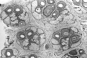 Figure 1. Transverse histologic section of adult male scalp at the level of the sebaceous glands showing 2, 3 and 4-hair follicular units (hematoxylin-eosin, X40)
THE LOGIC OF TRANSPLANTING INDIVIDUAL FOLLICULAR UNITS
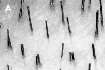
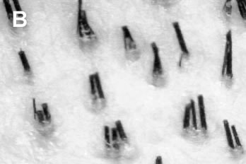
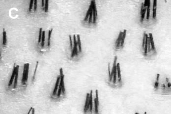
That scalp hair grows in follicular units, rather than individually, is most easily observed by densitometry, a simple technique whereby scalp hair is clipped to approximately 1mm in length and then observed via magnification in a 10mm field22. What is strikingly obvious when one examines the scalp by this method, is that follicular units are relatively compact, but are surrounded by substantial amounts of non-hair bearing skin. The actual proportion of non-hair bearing skin is probably on the order of 50%,16 so that its inclusion in the dissection will have a substantial effect upon the outcome of the hair restoration surgery. When multiple follicular units are used, and the skin is included, these effects may be profound.
To illustrate this point, use any of the “videografts” in figures 2 and draw a circle around a single follicular unit, and then draw a circle encompassing two units, then three etc. What one observes is that, as single follicular units are combined to form larger groups, the total volume of tissue included is not additive, but geometric.
When the actual transplant is performed, two additional factors act to compound the effects of this increased volume. The first is that the donor and recipient sites are not always a perfect match for one another. In many ways, transplanting skin from the back of the scalp to the front can be as different as using a graft from the inner thigh to fill in a defect on the lower leg. The reason is that bald scalp becomes atrophic over time, as the diminution of the follicular appendages are associated with a decrease in the other cutaneous elements.15
The other problem is that the transplantation of multiple follicular units, often requires recipient skin to be removed (via punch or laser) to allow this new volume of tissue to fit into the recipient site and/or to avoid unsightly compression of the newly transplanted grafts. In effect, richly vascular scalp, of maximum thickness, is transplanted into a somewhat atrophic recipient area, in which tissue is further removed to accommodate the graft. Not surprisingly, the results of this technique will often look unnatural!
The great benefit of using individual follicular units is that the wound size can be kept to a minimum, while at the same time maximizing the amount of hair that can be placed into it. Having the flexibility to place up to 4 hairs in a tiny recipient site has important implications for the design and overall cosmetic impact of the hair transplant surgery. It is a major advantage that follicular unit transplantation has over extensive micrografting in minimizing or eliminating the “see through” look that is so characteristic of the latter procedure.
THE LOGIC OF KEEPING RECIPIENT SITES SMALL
The importance of minimizing the wound size in any surgical procedure can not be over emphasized and hair transplantation is no exception. The effects of recipient wounding are felt at many levels. Large wounds can lacerate blood vessels and although the blood supply of the scalp is extensively collateralized, any damage to these vessels will have an impact on local tissue perfusion. An equally important issue is to minimize the disruption of the microcirculation. This is an unavoidable aspect of all scalp surgery, regardless of the size or depth of the wounds, but keeping this disruption to a minimum is a crucial part of the surgery. This is especially important when transplanting grafts in large quantities. The compact follicular unit is, of course, the ideal way to permit the use of the smallest possible recipient site, and has made the transplantation of large numbers of grafts technically feasible.9
Clearly, excision (removing tissue via a punch or laser) causes more damage to tissue then an incision (slit), but it is important to stress that all the parameters affecting recipient wounds have not been determined. As such, there are no absolute guidelines as to the ideal number, or densities of grafts, that can be used and still ensure maximum growth. The practitioner must rely on his clinical judgement in this regard, and it is suggested that one be conservative until one has significant clinical experience with the close placement of large numbers of grafts. In addition, there are a host of systemic and local factors that should be taken into account when planning the number and spacing of the recipient sites, regardless of their size.7
Another important advantage of the small wound is a factor that can be referred to as the “snug fit.” Unlike the punch, which destroys recipient connective tissue, a small incision, made with a needle, retains the basic elasticity of the recipient site. When a properly fitted graft is inserted, the recipient site will then hold it snugly in place. This “snug fit” has several advantages. During surgery, it minimizes popping, and the need for the sometimes-traumatic re-insertion of grafts. After the procedure, it ensures maximum contact of the implant with the surrounding tissue, so that oxygenation can be quickly re-established. In addition, by eliminating dead space, there is less coagulum formed, and wound healing is facilitated.
Since oxygen reaches the follicle by simple diffusion, its ability to do so is a function of tissue mass. Unlike larger grafts whose centers can become hypoxic, the slender follicular unit presents little barrier to this diffusion, thus ensuring uniform oxygenation.
It is important to note that when using larger grafts, either round or linear, compression is an undesirable consequence, and may result in a tufted appearance. In contrast, when transplanting follicular units, there are no adverse cosmetic effects of compression, since follicular units are already tightly compacted structures.
Another aspect of wound healing is the concept of “memory.” All of us who routinely perform cutaneous surgery, understand the advantage of wounds healing by primary intention. When tissue is removed by a punch, or destroyed by laser, the resulting defect heals by secondary intention. One can justifiably argue that when a graft is placed in the defect, the area doesn’t need to granulate in. However, because the underlying defect is still present, the wound invariably causes more scarring than when a simple incision is made (thus the term “memory”). This is readily evidenced in the scarred skin around the healed punch or laser sites. Although, not always visible, this tissue has lost its resiliency and cannot support the same density of grafts in subsequent procedures.
Large wounds cause a host of other cosmetic problems including dimpling, pigmentary alteration, depression or elevation of the grafts, or a thinned, atrophic look. The key to a natural appearing hair transplant is to have the hair emerge from perfectly normal skin. The only way to ensure this is to keep the recipient wounds small.
THE LOGIC OF CREATING SITES WITH COLD STEEL
In the public’s mind, no single word in medicine evokes a stronger image of “state-of the art” than the word “Laser,” and “Laser Hair Transplantation” is no exception.6 But, when the image begins to fade, and we examine its actions logically, we see that not only is the laser inappropriate for follicular unit transplantation, but that it is actually detrimental.
Lasers are used in hair transplantation to create recipient sites. In contrast to other fields of medicine where its properties of selective photo-thermolysis play a role, in hair transplantation the role is purely destructive. That lasers can create a hole with little surrounding thermal injury is little consolation to the surgeon who would prefer to have none. And the claim of the newest lasers, that they can make a recipient site with no thermal burn at all, is well and good, but it is missing the whole point. That point is that no matter how precise the laser is, it is still making a hole by removing tissue, and is, therefore, a throwback to the old punch technique.
Just to remind the reader, removing tissue destroys blood vessels and collagen, weakens the elastic support, increases the coagulum, decreases perfusion, and retards healing. Essentially, the laser “loosens” the “snug fit” that is such a benefit in follicular unit transplantation. If one merely wants to create a slit, which supposedly looks more natural than a hole, then lasers will do just fine. If one needs to remove tissue, to make room for a large graft, or prevent compression, then lasers may be the tools of choice. And, if one is more concerned that blood will cloud the view during surgery, rather than nourish the implants afterwards, then the laser should be given a try. But, if one wants to maximize the growth of follicular units, and keep recipient wounds to a minimum, then the beam should be pointed the other way.
THE LOGIC FOR TRANSPLANTING FOLLICULAR UNITS IN LARGE SESSIONS
Although larger sessions are made possible by the ability of follicular units to fit into very small recipient sites, and to minimize wounding, the next logical question to ask is “What is the actual advantage of performing these large sessions?” After all, they are time consuming, require a larger staff, and are more expensive for the patient (at least at the outset).
There are a number of very important reasons to transplant in large sessions. Some of them are specifically related to the use of follicular units, and some to hair transplantation in general, but all significantly affect our patient’s wellbeing. They may be summarized as follows:
• Social reasons
• Planning for telogen effluvium
• Economizing the donor supply
• Enhancing the complexion of the follicular units
The social implications of the surgery are uncommonly discussed at medical meetings, but are in the forefront of almost every balding patient’s mind. Putting aside anatomic, physiologic and technical issues for the moment, it is important to emphasize the practical reasons to strive toward large sessions. The specific events that bring a balding patient to the doctor for hair loss will vary, but the common denominator of those seeking hair restoration is to improve their appearance, and (although generally unspoken), to improve the quality of their life, be it personal, professional, or social.
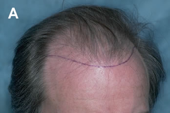
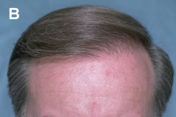
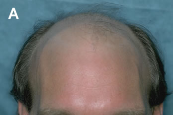
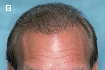
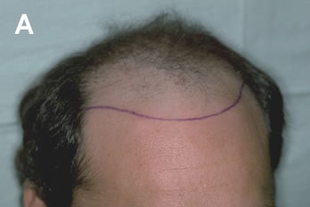
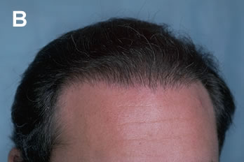
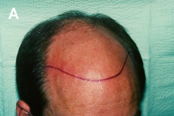
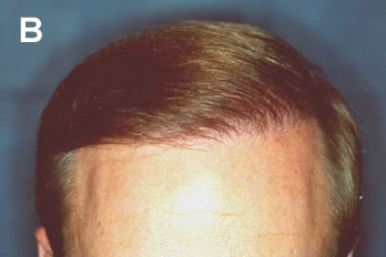
There is probably no better way for a surgeon to undermine this goal than to subject an already self conscious patient to a protracted course of small, incomplete procedures. Until the transplant is cosmetically acceptable, the disruptions from the scheduling of multiple surgeries, the limitations in activity, and the concern about their discovery, can place a patient’s life “on hold.” It should therefore be incumbent upon the physician to accomplish their objectives as quickly as possible. Figures 3 and 4 show what is possible using follicular units in large numbers in just one session, and figures 5 and 6 show what is possible in two sessions. The important point is that even if one or two transplant sessions does not accomplish all of a patients goals, he still can continue with normal activities while awaiting subsequent procedures.
Telogen Effluvium
Balding is a progressive process by which full-thickness terminal hairs gradually decrease in length and diameter in a process called miniaturization. This is a consequence of both the shortening of the anagen (growing) phase of the hair cycle and the diminution of the germinative elements in the follicle. Miniaturization is a universal aspect of androgenetic alopecia and accounts for most of the early cosmetic changes in hair loss. In other words, early in balding, the “thinning” that one notes is really due to thinning (i.e. miniaturization) of the hair shafts, rather than the actual loss of hair itself.9
Regardless of the technique, an inevitable aspect of hair transplant surgery is that the patient’s existing hair in, and around, the transplanted area has a chance of being shed as a result of the procedure. The hair that is at greatest risk of being lost is the hair that has already begun the process of miniaturization and, if this hair is at, or near the end of its normal life span, it may not return.
Often this shedding is mild and insignificant, but at times is can be substantial enough to leave the patient with a thinner look after the procedure than before he started. The reason is that in some patients (especially those that are younger and in very active stages of hair loss) large amounts of hair can be undergoing this process of miniaturization. Identifying those patients especially at risk, educating all patients that this process can occur, and planning for it surgically are thus integral parts of hair transplantation.12
The following is how one plans for this surgically:
1. Defer transplanting patients who are very early in the balding process, i.e. those who are content with the way they look now but are more concerned about future hair loss. A good rule of thumb is to wait unit the patient needs a minimum of approximately 600-800 follicular units before considering surgery. Often medical therapy, rather than surgery, would be appropriate for these patients.
2. When considering surgery, define the boundaries to be transplanted via densitometry as well as by gross visual inspection, such as densitometry, is a more sensitive indicator of miniaturization.
3. Transplant through (rather than around) an area that is highly miniaturized, since it is likely that this area will be lost by the time the transplant has grown in. Two examples of this would be a “forelock” composed of wispy miniaturized, rather than strong terminal hair or the “bridge” of a Norwood class 5 patient that is beginning to break down.
4. Plan to use enough follicular units so that, if possible, the volume of transplanted hair is greater than the volume of hair that will likely be lost from telogen effluvium. Remember, we are never replacing “hair for hair” in the surgery. We are, in effect, replacing a large number of fine, miniaturized hairs, with a much smaller amount of permanent, full-thickness terminal hairs.
In areas, of extensive miniaturization, it may be appropriate to transplant follicular units in the same density as if the area was totally bald.
Economizing The Donor Supply
As we mentioned in the introduction, the concern over the donor supply being finite is a rather recent development in hair restoration surgery, but since it is the ultimate limiting factor in all transplantation surgery, every possible effort should be made to insure the maximum yield. We would briefly like to review the logic of using large sessions with regard to the surgical wound. The importance of proper harvesting techniques and precise follicular dissection in ensuring maximum donor yield will be covered in later sections.
The donor supply is more sensitive to donor density than one might think. In fact, for every unit change in donor density, there is a 2-fold change in the amount of movable hair.9 Although not immediately obvious, the logic of this is illustrated in table 1. As will be discussed in the section “A Mathematical Look at Balding,” a person may loose 50% of his/her hair volume before it is clinically noticeable. Although we commonly think of this in terms of the balding scalp, it applies to the permanent zone as well.5 Therefore, in the average person with a density of 1 follicular unit/mm2 (2 hairs/mm2), the follicular unit density can be reduced to approximately 0.5 units/mm2 (1hair/mm2), before the donor area appears too thin. In those with high hair density, a greater percentage may be removed (see table 1). Therefore, a patient with a hair density of 2.5 hairs/mm2 would have 50% more movable hair than the average patient with a hair density of 2.0 hairs/mm2, although his hair density was only 25% more. The amount of movable hair will also depend upon other characteristics of the patient’s follicular units (see section “Characteristics of the Follicular Unit”) and upon his scalp dimensions and laxity.
The density will obviously be affected by each hair transplant. If a person has a hair transplant procedure(s) that decreases his donor density by 25%, then half of his movable hair will be exhausted, since his follicular unit density will be reduced to 0.75 units/mm2 (1.5 hairs/mm2). If that same patient, began with 25% less hair density i.e. 1.5 hairs/mm2 (remember, the follicular unit density is constant and would still be 1unit/mm2), then the same transplant(s) would reduce the follicular unit density to 0.75 units/mm2 and would leave a hair density of 1 hair/mm2 (0.75 units/mm2 x 1.5 hairs/unit), too thin to permit further transplantation. These numbers serve to underscore the importance of trying to conserve donor hair in every aspect of the procedure.
Table 1. THEORETICAL EFFECTS OF CHANGES IN DONOR DENSITY ON TRANSPLANTABLE HAIR*

** Although the patient with a donor density of 1.5 hairs/mm2 has ½ the available follicular unit grafts as a patient with a density of 3.0 hairs/mm2 (4,167 grafts Vs 8,333 grafts), each of his grafts, on the average, have only ½ the hair content of the patient with the density of 3.0, so that his transplant will appear only ¼th as full. (4,167 grafts averaging 1.5 hairs per graft Vs 8,333 grafts averaging 3 hairs per graft).
Regardless of how impeccable the surgical technique, each time an incision is made in the donor area, and each time sutures are placed, hair follicles are damaged or destroyed. This damage can be minimized (but not eliminated), by keeping the sutures very close to the wound edges so that they don’t encompass much hair. In subsequent procedures the damage can be reduced by using the previous scar as the upper or lower boarder of the new excision. In this way the amount of distortion and possible damage to existing hair is limited to only one free edge. Some physicians advocate the use of staples, feeling that this method of closure is quick, causes less trauma to the skin, and produces less damage to surrounding hair follicles (resulting in a superior scar). Others feel that staples produce unnecessary post-op discomfort and actually produce greater scarring due to the less controlled approximation of the wound edges. Studies still need to be performed to compare these two techniques and provide a definitive answer.
There are other more subtle effects of the surgery. In all healing, even with primary intention closures, collagen is laid down and reorganized. This distorts the direction of the hair follicles and increases the risk of transection in subsequent procedures. In addition, the fibrosis makes the scalp less mobile for subsequent surgeries, thus decreasing the amount of additional donor tissue that can be harvested. It should be clear that each time there is hair transplant surgery these factors come into play, so that transplanting in large sessions, which minimizes the total number of individual procedures, will conserve on total donor hair.
Complexion of Follicular Units
A final issue regarding the use of large sessions, is their ability to enhance the complexion of the follicular units generated from the donor strip. The logic behind this is very straightforward. In follicular unit transplantation the numbers of grafts present in any given size donor strip is determined by nature, since each graft represents one follicular unit. In contrast, in mini-micrografting techniques, the numbers are determined by the surgical team who cut the grafts “to size” depending upon how many of each size the surgeon feels are needed. For example if the “mini-micrografter” needed 200 single hair-grafts, he might divide up 100 two-hair grafts to produce 200 ones. If one felt he needed 100 4-hair grafts, he might combine 200 two-hair grafts to satisfy his needs. As we have discussed in earlier sections, this is strictly taboo in follicular unit transplantation, since the splitting of units risks damage and poor growth, and the combining of units produces unnecessarily large wounds and results that are not totally natural.
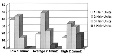
It follows that if we are to use only the naturally occurring individual units we are then limited by their normal distribution in the scalp and with larger sessions greater numbers of each type of unit will be generated. For example, in a scalp of average hair density (2.1 hairs/mm2), a donor strip of 1 cm x 20 cm would contain approximately 2,000 follicular units of the following distribution:
400 1 hair implants
1000 2 hair implants
500 3 hair implants
100 4 hair implants
In the same patient, a 5 cm strip of the same width would obviously contain 500 follicular units in similar proportions yielding:
100 1 hair implants
250 2 hair implants
125 3 hair implants
25 4 hair implants
In the average patient, it takes approximately 250 single hairs to create the soft transition zone of the frontal hairline, so in the smaller procedure the number of single hair grafts would be inadequate if one wanted to complete the procedure in one session. At the other end of the spectrum, one might need 500 three- and four-hair grafts placed in the “forelock” part of the scalp to give the patient a full, rather than diffusely thin look frontal. The smaller strip would only generate 250 of the larger three and four hair grafts, an inadequate number for this purpose. Clearly, then the logic of using larger procedures is that they will offer the surgeon the greatest flexibility in designing the transplant without having to combine or split follicular units.
As can be seen in figure 7, the patient’s absolute hair density will greatly effect the proportion of each of the 1-, 2-, 3-, and 4-hair follicular units found in the scalp. In patient’s with low hair density, a substantial proportion of follicular units will contain only a single hair and therefore the 1-hair grafts needed to construct a frontal hairline will be plentiful. In patients with high density, the higher proportion of the larger 3- and 4-hair units will provide the “natural resources” to create significant fullness in certain areas. How the different size follicular units are utilized will greatly affect the cosmetic outcome of the transplant and deciding their density and distribution is an “art” in itself.14
THE LOGIC OF THE FOLLICULAR UNIT CONSTANT
One of the interesting aspects of transplanting with follicular units is that nature was kind in spacing them at approximately one per square millimeter. Not only does this make the math easy, but it makes estimating the donor harvest easy, and gives us a logical basis for planning the density and distribution of the grafts. The relative constancy of the follicular unit density has been observed after performing densitometry on thousands of patients,9 and has been observed histologically by Headington as early as 1982.13
The follicular unit density is not exactly 1/mm2, but it is close enough to this number in most Caucasian and Asian scalps that it can be extremely useful in the surgery. In is significantly less in the black races, averaging around 0.6/mm2, and will decrease in everyone’s scalp as one moves laterally from the densest part over the occiput, towards the temples (see table 2).
It also tends to remain relatively constant with age.9 Finally, it is important to differentiate follicular unit density which is relatively constant, from hair density which can vary significantly from 1.5 hairs/mm2 to 3, or more, hairs/mm2 in the general population.3
Table 2. RACIAL VARIATIONS IN THE FOLLICULAR UNIT

Once one realizes that the follicular unit density is relatively constant and that hair density varies, it follows that the number of hairs per follicular unit largely determines hair density. In other words, patients with high hair density have more hairs per follicular unit rather than follicular units spaced closer together, and those with low hair density have fewer hairs per follicular unit, rather than follicular units spread further apart. One can easily see this relationship in the three videografts shown in figure 2. The implications of this in hair transplantation are enormous and can be summarized as follows:
• Since the follicular unit density is relatively constant, the same number of follicular units should generally be used to cover a specific size bald area regardless of the hair density of the patient.
• With low hair density, using the same number and spacing of follicular units as in a patient with high density, will help to ensure that there is proper conservation of donor hair for the long-term.
• Hair density is a characteristic of the follicular unit specific to each individual, and together with hair shaft diameter, color and wave, will determine the cosmetic impact of the transplant.
Traditionally, hair restoration surgeons have “sold” the hair transplant procedure to patients by promising the high density of larger grafts. In reality, the results are determined by the hair characteristics of the patient, rather than by the promises of the physician. In a patient with low hair density (or poor hair characteristics), each follicular unit has less cosmetic value, so the results will appear less full. On the other hand, in patients with high hair density and greater hair shaft diameter, the same number of follicular units will provide fuller coverage. Since the follicular density in each patient’s donor area is approximately the same, if we try to give the patient with fine hair and low density a “thick” look by combining them, we will simply run out of hair. Not to mention that combining the units will also produce a pluggy, unnatural appearance.
For most patients, the limitations of the donor supply compared to the demands of the recipient area are such that trying to transplant hair in a way that approaches, or equals, the donor density will limit the ability to properly distribute the hair on the long-term. Fortunately, it takes a surprising little amount of hair to make a differences in the appearance of a bald individual. Even in those individuals with thinning hair, the addition of even limited amounts of healthy terminal hair can radically change a person’s appearance. Logic might question this assumption, but clinical observations in thousands of patients have proven, over and over again, that a limited amount of hair, properly distributed, can radically improve the appearance of a balding man.
A Mathematical Look at Balding
To put things in perspective, let’s look at some aspects of the balding process mathematically.7 The normal hair density is approximately 2 hairs/mm2 or one follicular group/ mm2. The average person can loose 50% of his hair population without being detectably thin. That means that one needs to restore only one follicular unit every 2 mm2 in the hairline for a person’s density to appear normal from a frontal view. In areas behind the hairline, where layering of the hair can add value, significantly less than 50% of the original density may suffice to produce fullness. For example, with modest styling considerations, significantly less than 1/8th of the original density can appear to look full if placed behind a well constructed hairline.
In a typical patient with 50,000 follicular units on his scalp, the permanent donor area represents approximately 25% of this total number, or 12,500 units, with the remaining 37,500 at risk to be lost. Of the 12,500 units in the donor area, approximately half are available for harvesting (i.e. 6,250). We therefore have a total of 6,250 units to cover an area that originally had 37,500. We thus can replace only one sixth (6,250/37,500) of what we had to begin with. There are many creative ways to distribute the grafts so that the transplant has the appearance of being much fuller (see reference 7), but the point is that combining units to create more density is not one of them. This will only make the ratios worse.
For example, if we use only individual follicular units, the average spacing between units, once they have been transplanted into the recipient area, is six times further apart than their original spacing in the donor area (or six times further apart than in the pre-balding scalp). If we were to combine follicular units, i.e. combine three units into one, then the spacing increases to eighteen times as much. Visualizing the transplant process in this way, one can easily see that there is no logic in combining grafts to give more density. It only results in larger spaces, but never more hair. Fortunately, the patient with the thin looking donor area, will look appropriately balanced and natural with a thin looking transplant. The surgeon should promise no more.
Now that we realize that we can’t combine grafts to produce more fullness, how can we use the follicular constant to design the transplant and maximize the cosmetic impact? The issue is always one of long-term planning. Unfortunately, the patient doesn’t usually present with the final balding pattern. Therefore, when transplanting a patient early on, the density and distribution must be similar to how we would have transplanted him if he were further along in the process. Thus, if a patient has temporal recession at age 25, we shouldn’t give him any more density in this area than we would if he were 45. If we do, then when he is forty-five he will look unnatural.
Here is when an understanding of the follicular density comes in handy. If we have only one sixth the follicular density overall to work with and we want to use ½ the donor density in a certain area (i.e. 3x the average), then we can only use 1/18 the donor density (1/3 the average) in another area (given that these areas are of equal size) or we will run out of hair. For example, if we plan to eventually replace 50% of the patient’s original density in the forelock area, then some other region of the scalp must “give.” This might be accomplished by transplanting less on top of the scalp or transplanting the crown very lightly, or not at all. In the example of the 25 year old above, if we decide that the final density of the lateral aspects of the frontal hairline should be only ½ the density of the central “forelock,” then once we achieve this density, we must resist transplanting additional hair in that area, or the long-term distribution will be inappropriate.
The same would apply to the early treatment of the crown. If a patient presents with only crown balding, but because of his density, age, or family history he is expected to be very bald, one must place a limit on how much hair should be placed in this area. For example, it we assume that when he is completely bald and totally transplanted, the crown should have a density that is no greater than 1/20 the density of the donor area, then that is all that should be placed from the outset. Too often, a young patient, with a small area of balding is “packed” with hair to approximate the surrounding density and then later on he is left with a distribution so unnatural it can not be repaired.
Procedures which Defy Logic: Scalp Reductions and Flaps
The logic in using the follicular constant applies equally to other forms of hair restoration surgery. When analyzed in this manner, it becomes clear why flaps and scalp reductions cause so many long-term cosmetic problems2. In a flap, follicular units are moved from the donor to the recipient area in a one to one ratio, i.e. in a density that is six times what we have just shown to be appropriate. As a result, the flap will consume vast amounts of the donor supply to treat a relatively small portion of the balding scalp, and produce a transplanted density that always looks unnaturally high.
In scalp reductions, the donor skin that is moved is actually being stretched, so the density transfer is somewhat less than with a flap. For example, if two inches of bald scalp are removed from a series of scalp reductions (after any stretch-back has occurred), and three inches of donor fringe from each side have participated in this movement, then, in effect, six inches of hair-bearing scalp have been distributed over an area of eight inches. Assuming that the distribution is relatively uniform, the previously bald scalp now has a follicular density of approximately 75% of the donor density (6 divided by 8). On first blush one might think that this is a miracle. Especially since scalp reductions are presented as a technique which “…effectively conserves a significant donor area for future use.”, 23 we seemingly have gotten something for nothing. When viewed in the context of our previous discussion, where the crown was only being transplanted to 1/20 of the donor density to conserve hair for future transplants in the front, one wonders where all of this hair is suddenly coming from.
Remember we said that approximately one half of the donor supply could be used for transplantation before it appeared too thin? Well, we have just used up half of that 50% with the scalp reduction. In other words, if we can normally remove donor hair so that a follicular density of 1 unit/mm2 may be reduced to 0.5 follicular units/mm2 before it appears too thin, then our scalp reductions have already taken us to 0.75 units/mm2 or half way there (see preceding paragraph). And what have we gained by using up half of our donor supply? We’ve covered only two inches of bald area in the back of the head, thinned the scalp, and altered the normal hair direction. Furthermore, additional donor hair will be needed to cover the resulting scalp reduction scars and additional hair will be needed to address any new cosmetic problems as the balding in the crown progresses. In sum, scalp reductions have the unfortunate effect of simultaneously decreasing donor density and scalp laxity, and thus limiting the amount of hair available for the cosmetically important areas of the scalp.
Characteristics of the Follicular Unit
Before leaving this section, we just want to stress that when considering the cosmetic impact of the hair restoration procedure, it is important to consider all of the patient’s hair characteristics, as they can be of equal, or even greater importance, than the absolute number of hairs. For example, a close match of hair color and skin color will significantly contribute to the appearance of fullness, as will wavy hair. The follicular unit can actually be characterized by the following features:
• Hairs per follicular unit
• Hair shaft diameter
• Hair color
• Texture (wave, curl, kink)
• Other factors (emergent angle, static, oiliness, sheen, etc.)
It would seem logical to assume that the number of transplanted hairs is the major determinant in the cosmetic impact of the transplant. In reality, hair shaft diameter plays a more significant role than the absolute number of hairs. Coarse hair can be over twice the diameter of fine hair, so that when the area (~r2) of the hair shafts are compared, coarse hair has more than five times the cross-sectional area (and thus over 5 times the cosmetic value) of fine hair.3 If we compare this variance in hair shaft size to the natural variation in hair density, we can see that the impact of the hair shaft diameter (and thus volume) is over 2 ½ times as significant as the absolute number of hairs. Table 3 shows the range of hair density and hair shaft diameter commonly seen in the population of patients that are candidates for hair transplantation surgery and summarizes the relationship between the two. After reviewing this data, it should be apparent that an understanding of both the aesthetic and mathematical elements of transplantation are needed to predict the outcome of hair transplant surgery.
Table 3. RELATIVE SIGNIFICANCE OF HAIR DENSITY AND HAIR SHAFT DIAMETER

THE LOGIC OF SINGLE STRIP HARVESTING
The use of the multi-bladed knife is incompatible with follicular unit transplantation. When one remembers follicular unit anatomy and compares it to the construction of the knife, the reason should be obvious. The multi-bladed knife has blade spacing that generally range from 1.5 to 3mm. When these blades pass through a donor area that has follicular units randomly distributed at 1/mm2, few follicular units will be left unscathed. Clearly, one pass of the multi-bladed knife will break up many of the naturally occurring follicular units even before they leave the scalp and immediately reduce the follicular transplant procedure to one of mini-micrografts “cut to size.”8
The “lure” of the multi-bladed knife is that it quickly generates fine strips of tissue that can easily be dissected into smaller pieces, and the finer the strips, the easier the dissection. But, besides destroying the integrity of the follicular unit, the multi-bladed knife also causes transection to the follicles themselves, and the finer the strips the worse the transection. As was discussed in the first section, the multi-bladed knife is a form of “blind harvesting” that makes all of its incisions in a horizontal plane where the angle of the emerging hair is the most acute and the incisions can cause the most damage. Another issue complicating the harvesting is that follicular units actually transverse though the skin in a slightly curved path since the bulbs are random in the fat and “gathered” into bundles in the dermis.1 Regardless of the instrument, the initial incision is always going to be relatively “blind” so it makes the most sense to remove the strip with as few incisions as possible, and then perform all further cutting under direct visualization. This is the logic behind single strip harvesting.
It is argued by some, that a free hand ellipse is the preferred method for removing the strip, so that the cutting of each wound edge can be controlled separately (the upper edge should always be cut first). It is argued by others that two parallel blades offer more stability and avoids the problem of cutting through a mobile and partially distorted second edge (due to the greater contraction of the dermis relative to the epidermis and fat). Regardless of the personal preference of the surgeon, the concept is the same. Single strip harvesting is the best way to minimize transection, and the only way to provide adequate tissue for follicular unit transplantation.
THE LOGIC OF MICROSCOPIC DISSECTION
There is probably no other aspect of follicular unit transplantation that has generated more controversy than the use of the microscope. Fortunately, in no other area is the logic is more straightforward. Stereo-microscopic dissection was introduced into the field of hair transplantation by Dr. Bobby Limmer17 who recognized the logic of using this tool as early as 1987. Its full value and impact is only now first being appreciated. The following three statements summarize its use:
• You can only perform follicular unit transplantation if you have follicular units to transplant.
• In order to dissect intact individual follicular units, you must be able to see them clearly.
• Only the microscope allows their clear visualization in both normal and scarred skin, independent of the specific hair characteristics of color, hair shaft diameter, and curl.
Follicular dissection can logically be divided into two parts; the subdivision of the initial donor strip into smaller pieces and the further dissection of these pieces into individual follicular units. The first part of the procedure, the handling of the intact strip, has always been the most problematic. This is the main reason for the continued popularity of the multi-bladed knife as the multi-bladed knife, in effect bypasses the first part of the procedure by generating thin sections that can be laid on their sides. The thin sections, resting on their sides then have stability for further dissection and permit transillumination from back-lighting. The intact strip however, is difficult to stabilize and is too opaque for transillumination to be useful.
The dissecting microscope allows the strip to be divided into sections (or “slivers”) by actually going around follicular units leaving them intact. The dissecting stereo-microscope is able to accomplish this because of its high resolution (usually 5x more powerful than magnifying loops) and its intense halogen top-lighting that provides continuous illumination, as one dissects through the strip. Stability can easily be achieved by applying slight traction to the free end of the strip. The thin slivers are then laid on their sides and the microscopic dissection of the individual units is completed. With stereo-microscopic dissection, except for the outer edges of the ellipse, every aspect of the procedure is performed under direct visualization, so that follicular transection can be minimized and the follicular units maintained.
In a bilaterally controlled study,4 the dissecting microscope was compared to magnifying loops with transillumination, for the preparation of follicular unit grafts after the strip was divided into thin sections. The results showed that microscopic dissection produced a 17 % greater yield of hair as compared to magnifying loops with transillumination. This study showed an increase in both the yield of follicular unit grafts, as well as the total amount of hair. What is important to note is that this increase was observed when only the latter part of the dissecting procedure was studied i.e. after the strip has already been cut into sections. When complete microscopic dissection is used, the difference in yield is even more significant, and is probably on the order of an additional 5% to 10%.
THE LOGIC OF AUTOMATION
Although in concept, follicular unit transplantation may be the “ideal” transplant procedure, in its clinical application it poses special problems that have limited its widespread use.
• Follicular unit dissection is exacting and requires special skill.
• Follicular units are delicate and require special handling.
• Follicular unit transplantation is labor intensive and time-consuming.
One approach to these problems is to defend the status quo and rationalize the use of older techniques. The more logical solution is to solve them. The Rapid Fire Hair Implanter Carousel™* is a new instrument that has been designed to address these technological problems through automation. 21 The “Carousel” works by creating a recipient site with a specialized knife, and then “dragging” the graft into place as the knife is withdrawn. This “dragging” action is especially useful for small, delicate grafts, such as follicular units, as they normally tend to compress or bend when pushed. By combining site creation and graft placement into a single step, the extra time it would take for the grafts to be inserted separately is virtually eliminated. The cartridge, which holds 100 units at a time, is specially designed for procedures involving large numbers of grafts and may also be used in mini-micrografting techniques.
An interesting phenomena helps to explain why the Carousel places grafts into recipient sites so easily, while manual insertion is so problematic. The “finger” of the Carousel is able, in one motion, to insert the implant down to its final position in the skin. The surrounding tissues then apply a predominantly lateral force to the graft, holding it in place as the instrument is removed. In contrast, when grafts are inserted manually into a small site, the forceps allow only partial insertion on the first pass. While the forceps are being re-positioned, the vector of force on the graft is upward, and extrusion or popping occurs. Further attempts at insertion are clouded by the bloody field, and the implants, which were carefully grasped initially, are now grabbed across their growth centers and forced into the hole. This process causes crush injury and often irreversible damage. It is referred to in the literature as “H” or Human factor12, and is especially important when using small grafts that are more susceptible to mechanical trauma.
In all hair transplant procedures, economy of time is an essential element for success. Not only is a shorter procedure more practical for the staff and patient, but the grafts, subject to a relatively hypoxic environment once they have been removed from the body, are more quickly reunited with their oxygen supply. Although chilled holding solutions, greatly increase the survival of tissue outside the body, the sooner the grafts are re-inserted, the greater will be their chance of maximum growth. In large transplant sessions, the Carousel’s speed possibly represents its most important benefit.
Another source of injury that particularly affects small grafts is desiccation and warming. The grafts are at greatest risk while awaiting placement into the scalp, since at other times they can be held in chilled solutions. It has been recently shown that dried grafts are especially sensitive to mechanical trauma and will compound this form of injury (Gandleman M: Light and electron microscopic analysis of controlled crushing injury of micrografts. Presentation, ISHRS, Barcelona 1997). Warming will accelerate the effects of tissue hypoxia, as it speeds up the anaerobic metabolism. The enclosed cartridge of the Carousel helps to maintain a stable environment for the grafts from the time of dissection right until they are inserted into the scalp.
By reducing staffing requirements, operative time, and, most importantly, H-factor, automation appears to be a logical way of addressing many of the technical problems associated with the transplantation of large numbers of small grafts. Hopefully, this will allow a high-quality follicular unit transplantation procedure to be more easily performed by physicians and, as a result, be available to a greater number of our patients.
* Disclosure Statement: One of the authors of this article has a proprietary interest in Rapid Fire Hair Implanter Carousel™ manufactured by Apex Medical Products, LLC, Las Vegas, Nevada.
CONCLUSION
We have come full circle in our thought excursion through follicular unit transplantation. We began by showing some of the illogical events that led the field astray, and then how simple observation brought us back on track. We explained the logic of preserving the follicular unit and of using very small recipient sites. We saw the logic of using the follicular constant in the planning of the transplant and the advantage of performing it in large sessions. We saw the benefits of single strip harvesting and of microscopic dissection. Finally, we discussed problems intrinsic to the follicular unit transplantation procedure itself, and provided some logical ways to solve them.
Its seems that hair transplantation is a logical process after all! Why didn’t we notice that before?
References
1. Bernstein RM: A neighbor’s view of the “follicular family unit.” Hair Transplant Forum International 8:(3) No 3:23-35 1998.
2. Bernstein RM: Are scalp reductions still indicated? Hair Transplant Forum International 6:(3):12-13, 1996.
3. Bernstein RM: Measurements in hair restoration. Hair Transplant Forum International 8:(1):27, 1998.
4. Bernstein RM, Rassman WR: Dissecting microscope vs. magnifying loops with transillumination in the preparation of follicular unit grafts: A bilateral controlled study. Dermatologic Surgery 24: 875-880, 1998.
5. Bernstein RM, Rassman WR: Follicular Transplantation: Patient Evaluation and Surgical Planning. Dermatologic Surgery 23:771-784, 1997.
6. Bernstein RM, Rassman WR: Laser hair transplantation: Is it really state of the art? Lasers in Surgery and Medicine 19:233-235, 1996.
7. Bernstein RM, Rassman WR: The Aesthetics of Follicular Transplantation. Dermatologic Surgery 23:785-799, 1997.
8. Bernstein RM, Rassman WR, Seager D, et al.: Standardizing the classification and description of follicular unit transplantation and mini-micrografting techniques. Dermatologic Surgery 24:957-963, 1998.
9. Bernstein RM, Rassman WR, Szaniawski W, Halperin A: Follicular Transplantation. International Journal of Aesthetic and Restorative Surgery 3:119-132, 1995.
10. Bradshaw W: Quarter-grafts: a technique for mini-grafts. In Unger WP, Nordstrom REA, (eds): Hair Transplantation, ed 2, New York, Marcel Dekker, 1988.
11. Coiffman F: Injertos caudrados de cuero cabellerbo. Quinto, Ecquador: Primer Congresso Plastica, 1976.
11a. Gandleman M: Light and electron microscopic analysis of controlled crushing injury of micrographs.
Presented at the International Society of Hair Restoration Surgery. Barcelona, 1997.
12. Greco J: The H-factor in micrografting procedures. Hair Transplant Forum International 6:(4):8-9, 1996.
13. Headington JT: Transverse microscopic anatomy of the human scalp. Archives of Dermatology 120:449-456, 1984.
14. Hill T: Closure of the donor siste in hair transplantation by a cluster technique. Journal of Dermatologic Surgery and Oncology 6:190-191, 1980.
15. Lattanand A, Johnson WC: Male pattern alopecia: A histopathologic and histochemical study. Journal of Cutaneous Pathology 2:58-70, 1975.
16. Limmer B: Thoughts on the extensive micrografting technique in hair transplantation. Hair Transplant Forum International 6:(5):16-18, 1996.
17. Limmer BL: Elliptical donor stereoscopically assisted micrografting as an approach to further refinement in hair transplantation. Dermatologic Surgery 20:789-793, 1994.
18. Orentreich N: Autografts in alopecias and other selected dermatological conditions. Annals of the New York Academy of Sciences 83:463-479, 1959.
19. Pierce HE: An improved method of closure of donor sites in hair transplantation. Journal of Dermatologic Surgery and Oncology 5:475-476, 1979.
20. Pouteaux P: The use of small punches in hair-transplant surgery. Journal of Dermatologic Surgery and Oncology 6:12, 1980.
21. Rassman WR, Bernstein RM: Rapid fire hair implanter carousel: A new surgical instrument for the automation of hair transplantation. Dermatologic Surgery 24:623-627 1998.
22. Rassman WR, Pomerantz, MA: The art and science of minigrafting. International Journal of Aesthetic and Restorative Surgery 1:28-29, 1993.
23. Seager D: Binocular stereoscopic dissecting microscopes: should we use them? Hair Transplant Forum International 6:(4):2-5, 1996.
24. Stough DB: Hair Transplantation by the feathering zone technique: new tools for the nineties. American Journal of Cosmetic Surgery 9:243-8, 1992.
25. Tamura H: Hair grafting procedure. Japanese Journal of Dermatology and Venereology [Japanese] 52(2), 1943.
26. Uebel CO: The punctiform technique with the 1000-graft session. In Stough DB, Haber RS (eds): Hair Transplantation: Surgical and Medical. St. Louis, Mosby-Year Book, Inc. 1996, pp 172-177.
27. Unger WP: Usefulness of scalp reduction. In Unger WP, (ed): Hair Transplantation, ed 3, New York, Marcel Dekker, 1995.
28. Vallis CP: Surgical treatment of the receding hairline. Plastic and Reconstructive Surgery 33:247, 1964.


