One of the most important aspects of FUT Hair Transplant surgery is stereo-microscopic dissection. This allows follicular units to be removed from the donor strip without being broken up or damaged. During the dissection, it is critical that the whole follicular unit is kept intact as this will maximize its growth. Intact follicular units will also give the most fullness to the hair restoration, as they contain the full, natural complement of 1-4 hairs.

The average donor strip (above left) is approximately 1cm wide and of variable length, depending upon the number of follicular units that are needed for the hair restoration. In the average person’s scalp, there are approximately 90-100 follicular units per square cm of donor tissue, so a 2,000 graft hair transplant session would require a 1cm wide strip that is slightly over 20 cm in length.
The series of stereo-microscopes (above right) needed for the follicular unit dissection is located in the patient’s surgical room, so that graft dissection can occur simultaneously with other steps of the hair restoration and so that the hair transplantation can proceed seamlessly. Our highly-trained staff has years of experience assisting Dr. Bernstein in his pioneering Follicular Unit Transplantation procedure and you will not find a better team in the field of hair restoration. Such care is necessary to maximize graft survival by minimizing the risk of transection of follicles or mishandling.

The first step in stereo-microscopic dissection is a process called “slivering” (above left). In slivering, the dissector divides the long donor strip into smaller sections of approximately 2-2.5mm in width. The sectioning is accomplished by passing a scalpel blade through the spaces around follicular units, rather than merely cutting directly through the block of tissue. This technique generates smaller pieces of hair bearing tissue without breaking up follicular units and without transecting (cutting) hair follicles.
The dissector then isolates the individual follicular units from these small sections by trimming away excess dermis and fat. (Above right).

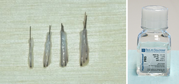
The photo (above left) shows 1-, 2-, 3-, and 4-hair follicular units. Hundreds of these individual units are sorted according to the numbers of hairs they contain.
They are then stored in HypoThermosol® (above right), an optimized hypothermic (low temperature) preservation medium. We use HypoThermosol because it contains macro-molecules that best mimic the intra-cellular fluids. When the grafts are chilled, this solution prevents small molecules, such as Na+, from entering the cells and causing damage. HypoThermosol® also contains two potent antioxidants, glutathione and a vitamin E analog. Together, they mitigate temperature-induced molecular cell stress that occurs during the chilling and re-warming of grafts. This results in significantly extended viability of the grafts while waiting to be placed back into the scalp.

Once the recipient sites have been created, a portion of the grafts are removed from the refrigerated storage and placed into Petri-dishes that sit on a wall-mounted placing stand at the head of the operating table (above left). The grafts are bathed in a HypoThermosol® solution (above right) at a constant temperature while waiting to be placed back into the patient’s scalp.


